What is Tricuspid Atresia?
Tricuspid atresia is a congenital heart defect characterized by the abnormal development or absence of the tricuspid valve.1 The tricuspid valve is in between the right atrium and right ventricle and ensures blood flows in the correct direction, from the atrium to the ventricle.2 In a healthy heart, deoxygenated blood would flow into the heart’s right atrium, pass through the tricuspid valve, and move into the right ventricle before entering the lungs for reoxygenation.2 In tricuspid atresia, there is a tissue in place of the tricuspid valve, which blocks adequate blood flow from the right atrium to the right ventricle. Consequently, blood cannot pass through to the lungs via the normal route.3 Tricuspid atresia is often accompanied by a ventricular septal defect (VSD) or atrial septal defect (ASD). VSD is a hole between the lower heart chambers (the ventricles), allowing blood to flow through the hole to the main lung artery. ASD is a hole in the upper heart chambers. Tricuspid atresia means that blood is forced through the hole into the left atrium, mixing with oxygen-rich blood that has returned from the lungs. This mix of blood is pumped through the body by the left ventricle, resulting in a lack of oxygen throughout the entire body.1 Another thing to note is that the right ventricle and pulmonary valve, the valve that exists between the right ventricle and the artery that takes deoxygenated blood to the lungs, typically becomes underdeveloped and abnormally small due to the blockage and lack of use.1,3

Missing tricuspid valve: the valve in between the RA and RV is missing, and instead, there is tissue which blocks adequate blood flow from the RA to RV. Blood returning to the heart via the right atrium must find an alternative route, typically through a hole between the right and left atria. Consequently, blood cannot pass through to the lungs via the normal route.
Ventricular septal defect: VSD is a hole between the lower heart chambers (the ventricles), which eventually allows blood to flow through the hole to the main lung artery so that it can be oxygenated.
There are three variations of tricuspid atresia:
-
The pulmonary artery and aorta are in the right place, but might have a hole in their ventricular wall (VSD) or a problem with their pulmonary valve.
-
The pulmonary artery and aorta are swapped and in the wrong place. There is also a VSD and/or an issue with the pulmonary valve.
-
The rarest type is when there are several problems with the position of the pulmonary artery, aorta, and the right and left ventricles
Symptoms
Tricuspid atresia’s symptoms can vary based on each patient and their specific defect. In patients with pulmonary obstruction (the narrowing or blockage of the airway, making it difficult to breath) other symptoms can include:4
cyanosis (blue coloring of lips, skin, nail beds)
shortness of breath
difficulties in feeding
fatigue
failure to thrive
Patients without pulmonary blockage can have less obvious symptoms than those with obstruction. Symptoms can include:3
heart failure
respiratory distress- rapid, shallow breathing that can sound like grunts
impaired growth
poor feeding
rapid breathing (tachypnea)
rapid heart rate (tachycardia)
enlarged liver (hepatomegaly)
Diagnosis
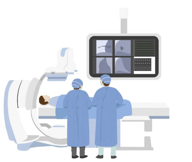
In terms of diagnosis, advancements in medical technology have allowed congenital defects to be diagnosed earlier, during pregnancy, through antenatal ultrasounds and fetal echocardiograms. If it is not diagnosed prenatally, it is usually diagnosed shortly after birth by means of:
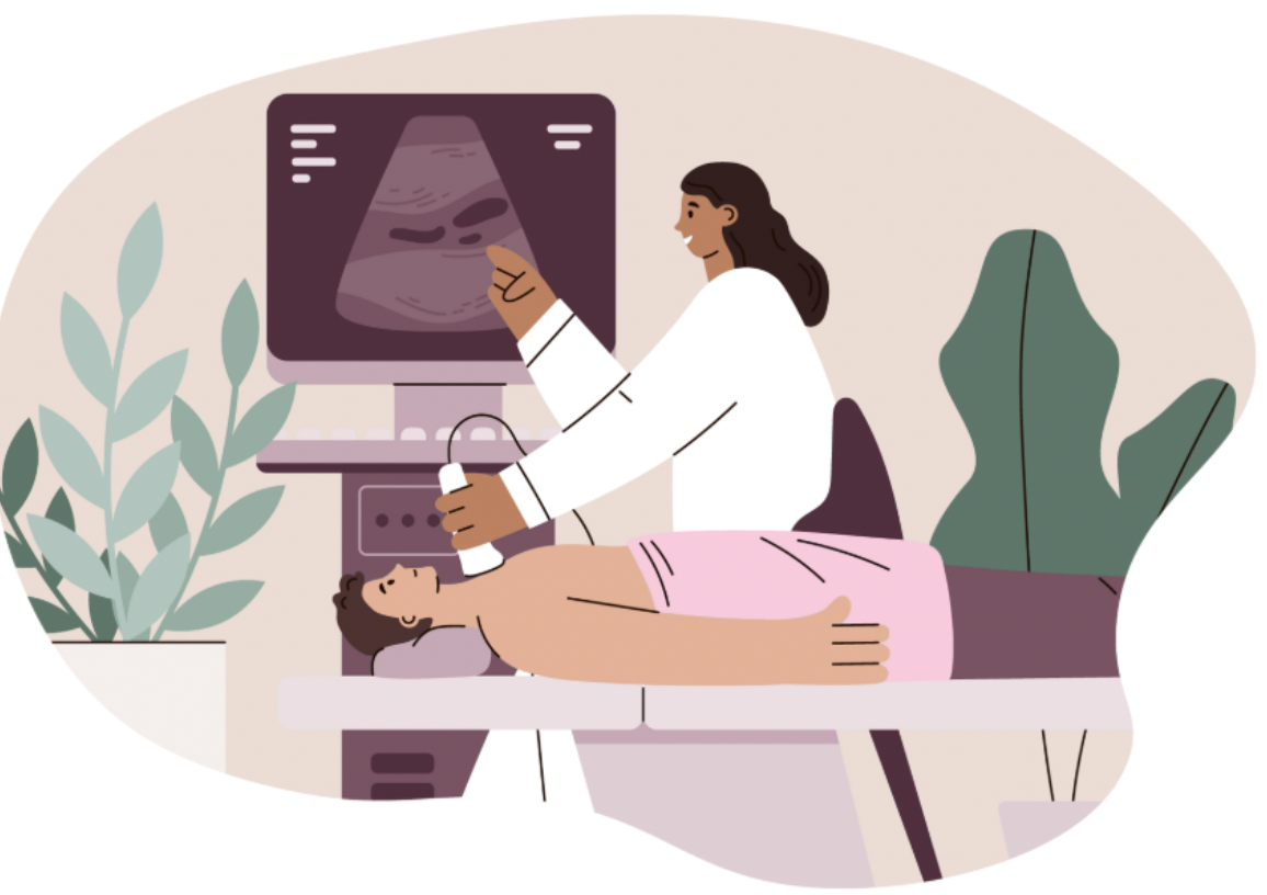
Ultrasound

Chest X-ray (CXR)
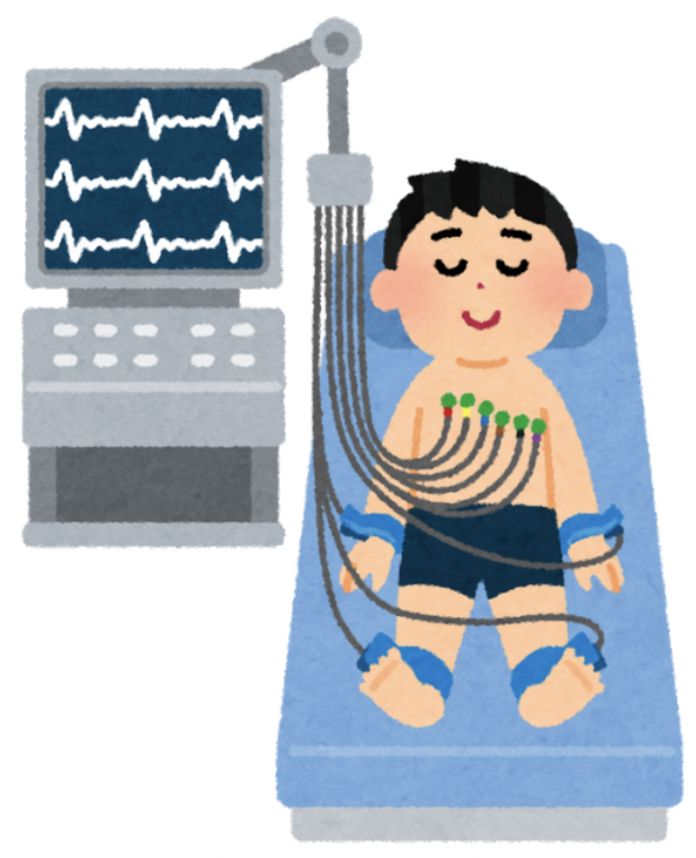
Electrocardiogram (EKG)
Cardiac Catheterization

Echocardiogram (Echo)
These tests detect the absence of the tricuspid valve, atrophy of the right ventricle (atrophy means that the tissue is decreasing in size and wasting away due to lack of function), blood flow (whether it is passing through the the valve or walls between the heart chambers) and other related defects.3
Treatments
Treatment for tricuspid atresia, as well as other single ventricle defects, is often done in stages. Once diagnosed, the initial goal is to stabilize the patient.4
Although it depends on the severity of the patient's condition, this usually involves admission to the neonatal intensive care unit (NICU) and initiation of a medicine called prostaglandin E1.2
Then, there are a series of surgeries including the Blalock-Taussig shunt, the Glenn procedure or hemi-Fontan procedure, and the Fontan procedure.1-5
Additional Resources
References:
1. John Hopkins. Tricuspid Atresia. Accessed December 7, 2024. https://www.hopkinsmedicine.org/health/conditions-and-diseases/tricuspid-atresia
2. Medline Plus. Tricuspid Atresia. Accessed December 7, 2024. https://medlineplus.gov/ency/article/001110.htm
3. Cleveland Clinic. Tricuspid Atresia. June 28, 2022. Accessed December 7, 2024. https://my.clevelandclinic.org/health/diseases/14789-tricuspid-atresia
4. Prashant K. Minocha; Maria S. Horenstein; Colin Phoon. Tricuspid Atresia. National Library of Medicine. January 6, 2024. Accessed December 7, 2024. https://www.ncbi.nlm.nih.gov/books/NBK554495/
5. Anoop S. Sumal BA, Harry Kyriacou BA, Ahmed M. H. A. M. Mostafa BA. Tricuspid atresia: Where are we now? Wiley Online Library. June 2, 2020. Accessed December 7, 2024. https://onlinelibrary.wiley.com/doi/10.1111/jocs.14673