What is Hypoplastic Left Heart Syndrome?
Hypoplastic Left Heart Syndrome (HLHS) is characterized by the underdevelopment of the left-sided structures of the heart, including the mitral valve, left ventricle, aortic valve, ascending aorta, and aortic arch.1 This underdevelopment can be caused by obstruction of the blood moving out of the left ventricle and into the aorta via the aortic valve, or obstruction of the tract where blood would enter the left ventricle from the left atrium.1 The most common cause of underdevelopment is aortic valve stenosis, the thickening and narrowing of the valve between the left ventricle, or main pumping chamber, and the aorta. If the left ventricle outflow is blocked, it increases the pressure the heart must overcome to contract and pump blood out, resulting in enlargement (sometimes referred to as hypertrophy) of the left ventricle muscle, increased LV dilation, and decreased muscular contraction of the left ventricle.2 The build up in blood in the left ventricle due to the lack of pumping ability can lead to a pressure increase in the left atrium, and blood can start to flow from the left atrium to the right atrium through the foramen ovale (this is the reverse of what we want to happen).1 As mentioned, the other cause of HLHS can be that the blood entering the left ventricle from the left atrium is obstructed. This can be caused by complete blockage or narrowing of the mitral valve (which lies between the left atrium and left ventricle) or a restrictive foramen ovale (which typically allows blood to flow from the right atrium to the left atrium). In both instances, reduced blood flow through the LV limits the growth and development of the left ventricle (referred to as hypoplasia).1
Underdeveloped aorta

Open ductus arteriosus.
Underdeveloped left side of the heart: significantly smaller than normal.
Symptoms:
HLHS’s symptoms can vary based on each patient and their specific defect. Symptoms of HLHS typically appear within 2-3 days. Symptoms can include:1-3
Blue or purple tint to lips, skin, and nails (cyanosis)
Difficulty breathing
Difficulty feeding
Sleepy or unresponsive (lethargy)
Weak pulses
Poor circulation (poor perfusion)
Fast heart rate (tachycardia)
Low blood pressure (hypotension)
Diagnosis
HLHS is typically diagnosed by prenatal ultrasounds.1 But, sometimes it is diagnosed hours or even days after birth.3 These babies are usually born at term and a heart murmur is not present. Their lack of symptoms for the first 24-48 hours is attributed to their ductus arteriosus still being open, allowing for oxygenated blood to travel from the pulmonary artery (the vessel delivering oxygenated blood from the lungs back to the heart) and the aorta (the main vessel that delivers blood to the rest of the body). It is often diagnosed by means of:1-4
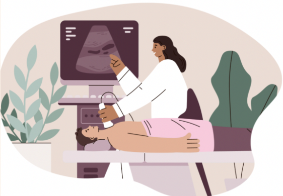
Echocardiogram (Echo)
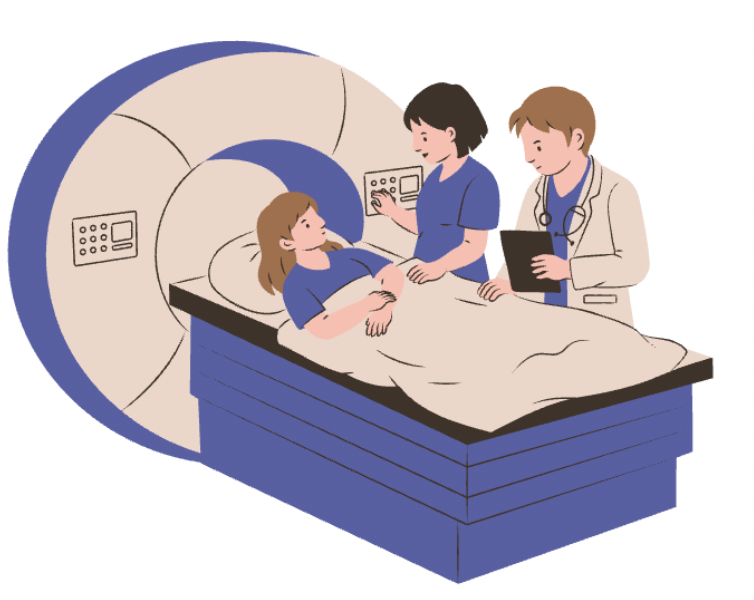
Cardiac MRI
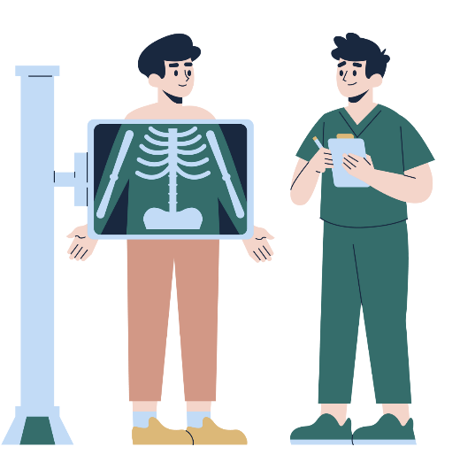
Chest X-ray (CXR)
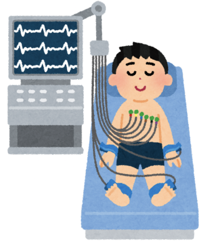
Electrocardiogram (EKG)
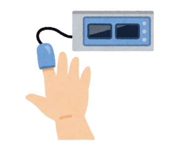
Pulse Oximetry
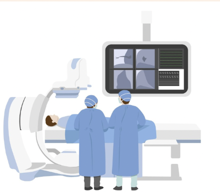
Cardiac Catheterization
Treatments
Treatment for tricuspid atresia, as well as other single ventricle defects, is often done in stages. Once diagnosed, the initial goal is to stabilize the patient. Although it depends on the severity of the patient's condition, this usually involves admission to the neonatal intensive care unit (NICU) and initiation of a medicine called prostaglandin E1.1 Prostaglandin E1 is given to keep the ductus arteriosus open (the ductus arteriosus is a short blood vessel that connects the fetal pulmonary artery to the aorta). Keeping this vessel open, which typically closes after birth, allows blood to flow from the pulmonary artery to the aorta and presents an alternate route for blood to receive oxygen. If there is a restrictive foramen ovale or intact atrial septum, the newborn will be taken to the cardiac catheterization lab to enlarge and decompress the left atrium.1 The patient will require open heart surgery to redirect the oxygen-rich blood and oxygen-poor blood. The series of three reconstructive operations that typically take place include the Norwood, Glenn, and Fontain procedures. In severe cases, heart transplantation may be necessary.1, 3, 4
Additional Resources
References
Stacy M. Kritzmire; Anne E. Cossu. Hypoplastic Left Heart Syndrome. National Library of Medicine. April 24, 2023. Accessed December 7, 2024. https://www.ncbi.nlm.nih.gov/books/NBK554576/
Anthony T Bejjani, Neil Wary, Mingxia Gu. Hypoplastic left heart syndrome (HLHS): molecular pathogenesis and emerging drug targets for cardiac repair and regeneration. National Library of Medicine. September 15, 2024. Accessed December 7, 2024. https://pmc.ncbi.nlm.nih.gov/articles/PMC8511342/#:~:text=Hypoplastic%20Left%20Heart%20Syndrome%20(HLHS)%20is%20a%20%E2%80%9Csingle%20ventricle,first%20year%20of%20life1.
Children’s Hospital of Philadelphia. Hypoplastic Left Heart Syndrome (HLHS). Accessed December 1, 2024. https://www.chop.edu/conditions-diseases/hypoplastic-left-heart-syndrome-hlhs
Roberts Gobergs, Elza Salputra, Ingūna Lubaua. Hypoplastic left heart syndrome: a review. National Library of Medicine. Accessed December 7, 2024. https://pmc.ncbi.nlm.nih.gov/articles/PMC5088741/