How a Healthy Heart Works
Structure and Function of an Adult Heart
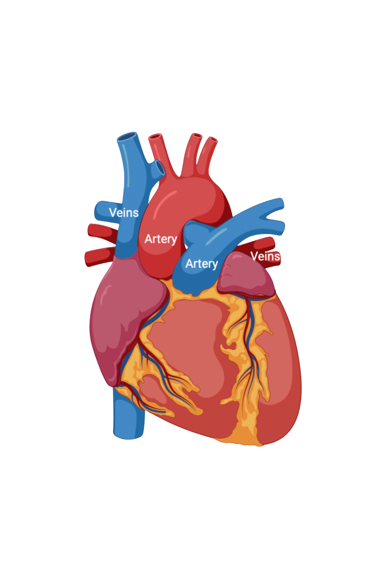
The heart’s main function is to move oxygenated blood throughout the body. Any blood vessels carrying blood to the heart is a vein, and any blood vessel carrying blood away from the heart is an artery.1 For example, the vessel carrying blood from the heart to the lungs is named the pulmonary artery, yet the vessel carrying blood from the lungs to the heart is named the pulmonary vein.

How Blood Flows Through The Heart
There are four main chambers of the heart: two atria (right and left) and two ventricles (right and left).2 Between each chamber and between a chamber and a vessel, there is a valve. The valves prevent blood from flowing in the wrong direction through the heart.3
Between the right atrium and right ventricle: tricuspid valve4
Between the right atrium and pulmonary artery: pulmonary valve4
Between the left atrium and left ventricle: bicuspid valve4
Between the left ventricle and the aorta: aortic valve4
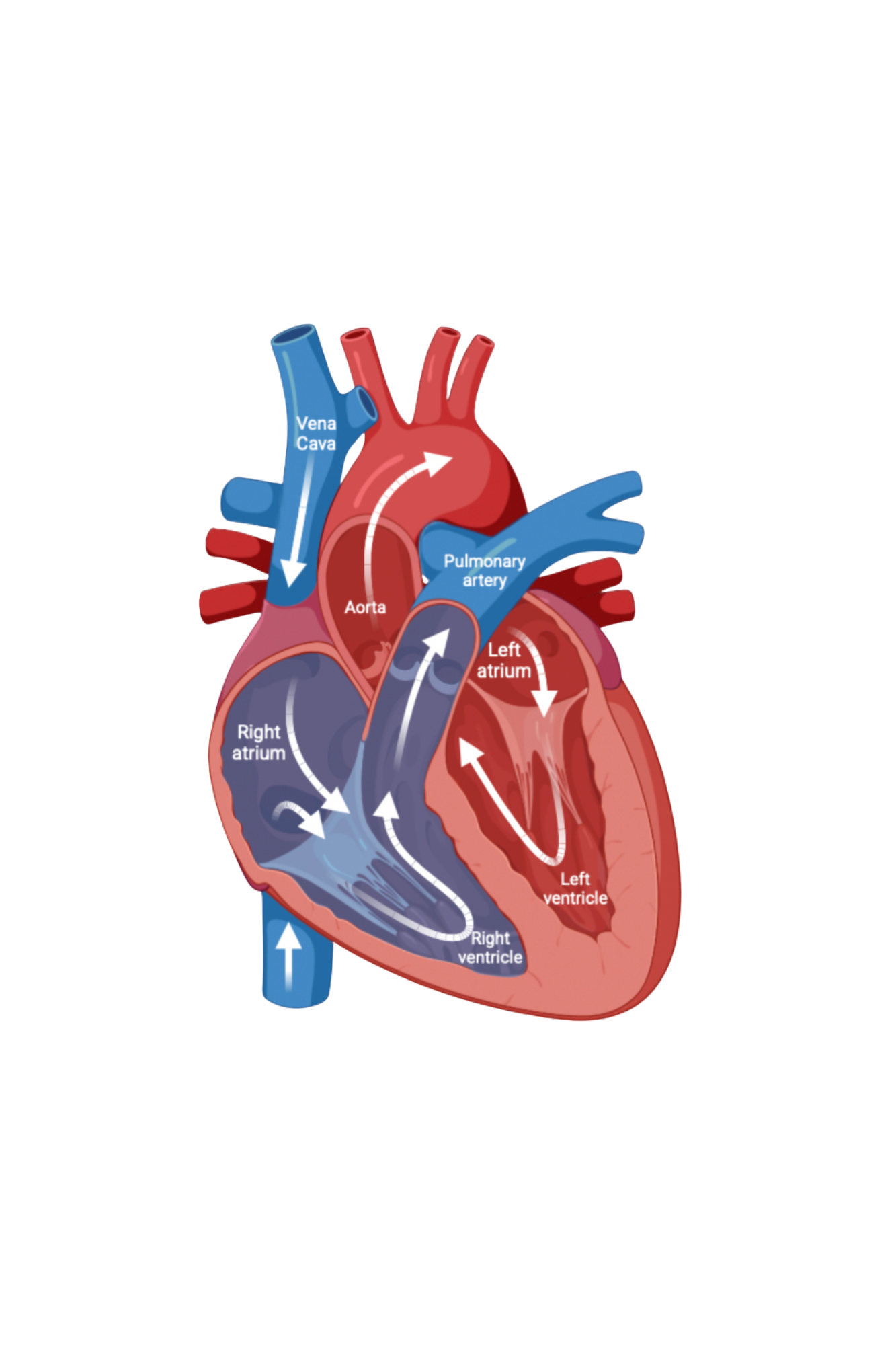
Oxygen-poor blood returns to the heart through two main veins: the superior vena cava (SVC) and inferior vena cava (IVC).5 The oxygen-poor blood enters the right side of the heart and flows into the right atrium (RA). The blood will continue to flow through the tricuspid valve (TV) and into the right ventricle (RV).4 The heart’s muscles contract (what you feel as a heart beat), and this forces the blood from the RV through the pulmonary valve into the pulmonary artery.5 During this contraction, due to the pressure build up, the tricuspid valve slams shut and prevents blood from flowing backwards into the RA. In the lungs, the blood will become reoxygenated as it receives oxygen and gets rid of carbon dioxide.2 The reoxygenated blood will then reenter the heart through the pulmonary veins into the left atrium (LA). The blood will flow through the mitral valve and into the left ventricle (LV).4 The LV is the most muscular chamber of the heart. This is because it must generate the high pressure required to pump blood through the whole body via the aorta.2 When the muscles in the heart contract, especially the muscles in the LV’s wall, blood is propelled through the aortic valve and into the aorta. The aorta is the main artery that is responsible for delivering oxygen-rich blood to the body.4
Whenever you feel a heartbeat that is the muscle in your heart wall contracting.1 The contraction decreases the volume in the chambers of your heart, and pushes the blood either to the lungs or to the rest of the body. Your heart has an electrical system that determines how fast your heart beats.2,5 There are two sounds typically heard when a healthy heart beats: “lub-DUB”. A typical heart first contracts to push blood from the atria into the ventricles (both sides of the heart do this at the same time). Once your atria have finished contracting, and as the ventricles begin to contract, the valves between atria and ventricles close. This generates the “lub” sound. Once your ventricles finish contracting, the valves between the aorta and the LV, and the pulmonary artery and the RV close. This generates the “dub” sound. Therefore, there are 2 contractions for every heartbeat – one that contracts both atria simultaneously, and one that contracts both ventricles simultaneously.
The Fetal Heart and Circulation
The fetal circulatory system works differently before and after birth. In the womb, to bypass the lungs and liver, organs that will not fully function until after birth, the fetus’s circulatory system uses three shunts.6
The fetus receives oxygen and nutrients from the mother’s blood through the placenta.6 The oxygen-rich blood flows through the umbilical vein and enters the inferior vena cava (one of the major vessels that brings blood back to the RA of the heart). The ductus venosus is used to bypass the liver at this stage. Once the blood is in the RA, it can either pass through the tricuspid valve and into the RV (which is what happens after birth), or it can pass through the hole between the atria called the foramen ovale. This means that blood in the atria can mix.
If blood goes through the right ventricle pathway and then onto the pulmonary artery, there is again another shunt present (the ductus arteriosus) that allows the blood to mix between the pulmonary artery and the aorta.6 This means that the blood bypasses the lungs (which are not inflated or working while the fetus is in the womb). However, once the baby is born and their lungs inflate, there is no longer any need to bypass the lungs. Therefore, mixing of oxygenated and deoxygenated blood through these shunts is no longer beneficial and makes the circulation less efficient. Oftentimes, these shunts will close shortly after birth, unless prostaglandins are provided.
-
The Foramen Ovale bypasses the lungs and moves the blood from the right atrium to the left atrium in the heart.
-
The ductus arteriosus directs blood from the pulmonary artery into the aorta and allows the blood to bypass the lungs.
-
The ductus venosus allows oxygenated blood from the mother’s placenta to bypass the liver and head straight to the heart
References
Cleveland Clinic. Heart. Cleveland Clinic. January 26, 2024. Accessed February 2, 2025. https://my.clevelandclinic.org/health/body/21704-heart
Children’s Hospital of Philadelphia. How the Normal Heart Works. Children’s Hospital of Philadelphia. Accessed February 2, 2025. https://www.chop.edu/centers-programs/cardiac-center/how-normal-heart-work
Mayo Clinic Staff. Atrioventricular canal defect. Mayo Clinic. September 13, 2022. Accessed February 2, 2025. https://www.mayoclinic.org/diseases-conditions/atrioventricular-canal-defect/symptoms-causes/syc-20361492
National Heart, Lung, and Blood Institute. How Blood Flows through the Heart. National Heart, Lung, and Blood Institute. January 24, 2022. Accessed February 2, 2025. https://www.nhlbi.nih.gov/health/heart/blood-flow
U.S. Centers for Disease Control and Prevention. Congenital Heart Defects (CHDs). U.S. Centers for Disease Control and Prevention. October 4, 2024. Accessed February 2, 2025. https://www.cdc.gov/heart-defects/how-the-heart-works/index.html
Stanford Medicine. Fetal Circulation. Stanford Medicine Children’s Health. https://www.stanfordchildrens.org/en/topic/default?id=fetal-circulation-90-P01790