
Cardiac MRI/CT 5,8
-
Magnetic Resonance Imaging (MRI) uses radio waves and magnets to create detailed images of the heart and blood vessels.
Computed Tomography (CT) uses x-rays from multiple different angles to depict an image of your heart.
Both CT and MRI are non-invasive procedures.
Results can be used to identify the structures of the heart and potential heart defects.
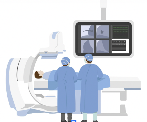
Cardiac Catheterization 6
-
A medical procedure where a flexible hollow tube (catheter) is inserted into a blood vessel (usually in the arm, groin, or neck) that is then threaded through the blood vessel and into the heart.
It can be used to treat or diagnose some heart conditions.
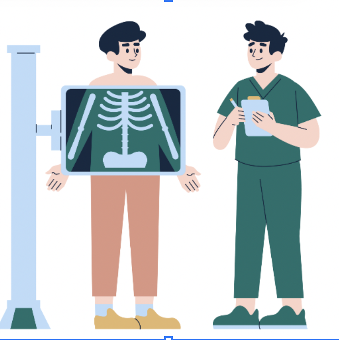
Chest x-ray:3
-
An imaging technique that uses x-rays to produce images of the heart, lungs, blood vessels, airways, and bones of the chest and spine.
A noninvasive imaging test that uses a small amount of radiation to produce an image of the chest.
Used to diagnose and monitor conditions affecting the structures inside the chest.
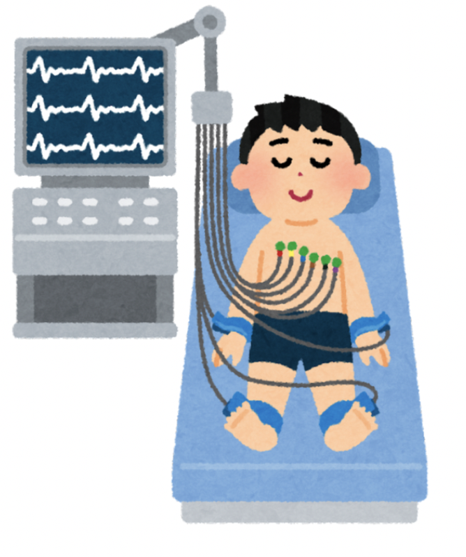
Electrocardiogram (EKG):1
-
Uses electrodes attached to your skin (normally in the chest, arms, or legs) to record electrical signals of the heart
Results are typically shown as a line graph.
An EKG can detail the patient’s heart rate, rhythm, timing of contractions, strength of contractions, and the size and position of the heart’s chambers
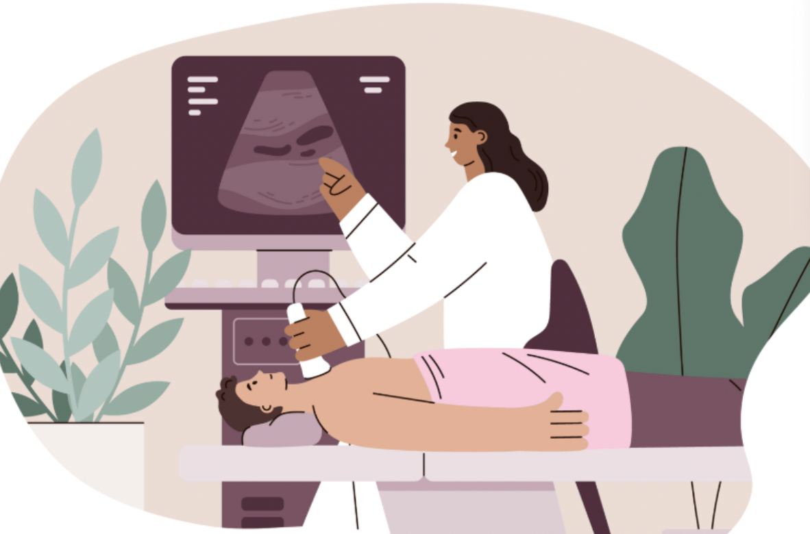
Echocardiogram:2
-
Also known as cardiac ultrasound.
An imaging technique that uses sound waves to create images of the heart and nearby blood vessels.
Shows how blood flows through the heart and heart valves.

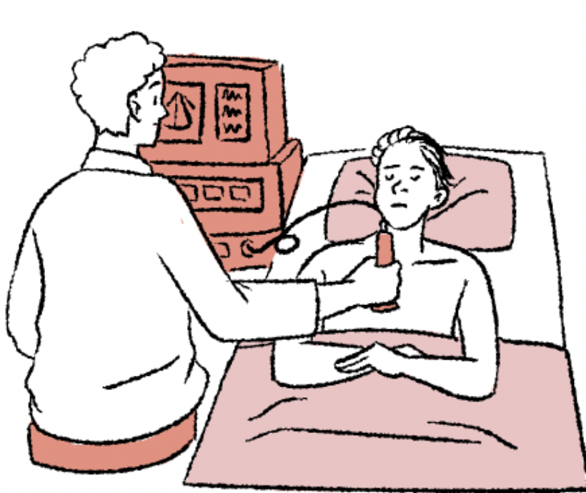
Exercise stress tests:7
-
A test where you wear an electrocardiogram whilst exercising on a treadmill or standardized bicycle
Measure your heart’s function under stress (4 - EA)
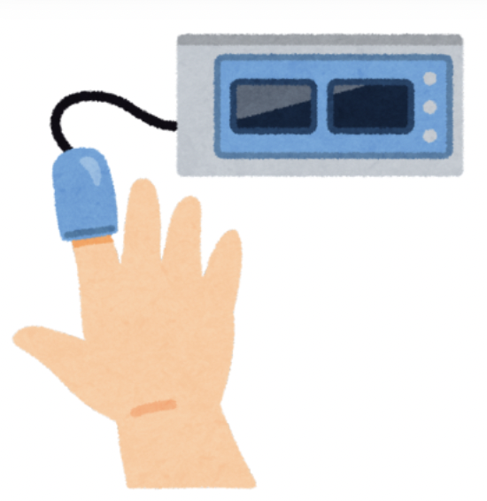
Pulse oximetry:4

-
This device measures the amount of oxygen in your blood.
The patient puts the device on the tip of their finger and the electronic device will measure the saturation of oxygen carried in your blood cells.
Holter monitor:7
-
Portable ECG device that records the heart's activity during daily activities(2)
Can be worn for 24-48 hours while the heart’s information is collected. It collects information from each heart beat including your heart’s rhythm and rate over long periods of time (9)
Ultrasound:
-
uses high-frequency sound waves to create real-time photos of internal organs and soft tissues
References:
Dr. Steven Reisman. 12-lead Electrocardiogram (EKG). New York Cardiac Diagnostic Center. March 5, 2023. Accessed December 21, 2024. https://newyorkcardiac.com/12-lead-electrocardiogram-ekgElectrocardiogram(EKG)
Mayo Clinic Staff. Echocardiogram. Mayo Clinic. Accessed December 21, 2024. https://www.mayoclinic.org/tests-procedures/echocardiogram/about/pac-20393856
Cleveland Clinic. Chest X-Ray. Cleveland Clinic. November 20, 2023. Accessed December 21, 2024. https://my.clevelandclinic.org/health/diagnostics/10228-chest-x-ray
American Lung Association. Pulse Oximetry. American Lung Association. Accessed December 21, 2024. https://www.lung.org/lung-health-diseases/lung-procedures-and-tests/pulse-oximetry
The American Heart Association. Cardiac Magnetic Resonance Imaging (MRI). The American Heart Association. April 13, 2023. Accessed December 21, 2024. https://www.heart.org/en/health-topics/heart-attack/diagnosing-a-heart-attack/magnetic-resonance-imaging-mri
Cleveland Clinic. Double Inlet Left Ventricle. Cleveland Clinic. October 27, 2022. Accessed December 21, 2025. https://my.clevelandclinic.org/health/diseases/14786-double-inlet-left-ventricle
Mayo Clinic Staff. Ebstein anomaly. Mayo Clinic. June 9, 2023. Accessed September 18, 2025. https://www.mayoclinic.org/diseases-conditions/ebsteins-anomaly/symptoms-causes/syc-20352127#:~:text=Ebstein%20anomaly%20is%20a%20rare%20heart%20problem%20that's%20present%20at,is%20called%20the%20tricuspid%20valve.
Cleveland Clinic. CT (Computed Tomography) Scan. Cleveland Clinic. Accessed July 16, 2025. https://my.clevelandclinic.org/health/diagnostics/4808-ct-computed-tomography-scan
Children’s Hospital of Philadelphia. Ebstein’s Anomaly of the Tricuspid Valve. Children’s Hospital of Philadelphia. Accessed January 18, 2025. https://www.google.com/url?q=https://www.chop.edu/conditions-diseases/ebstein-s-anomaly-tricuspid-valve&sa=D&source=editors&ust=1737258106456685&usg=AOvVaw20baziv9jr_TMDQPexaVuo