What is Pulmonary Atresia with Intact Ventricular Septum?
Pulmonary atresia is when there is obstructed flow from the right ventricle to the lungs due to the underdevelopment or abnormal development of the valve between the heart and the lungs (the pulmonary valve).1,2 This closure prevents blood from flowing from the right ventricle to the lungs and as a result, blood cannot flow to the lungs to become oxygenated.3 Pulmonary atresia with an intact ventricular septum means that while there is a lack of pulmonary valve development, there is no hole between the right and left ventricles. This means that the deoxygenated blood in the right ventricle does not mix with the oxygenated blood in the left ventricle. In patients without a ventricular septal defect (VSD), the right ventricle receives minimal blood flow before birth, resulting in underdevelopment and inability to function as a pumping chamber. Therefore, there is an underdeveloped right ventricle and only one functioning left ventricle.2,3 This can also cause further underdevelopment of the valve between the right atrium and the ventricle (tricuspid valve)2. Blood is redirected through a hole in the right atrium to the left atrium (known as the foramen ovale), and survival is dependent on the hole remaining open until a shunt is placed.4
Open ductus arteriosus:

Underdeveloped/abnormal pulmonary valve: the valve between the right ventricle and the pulmonary artery (going to the lungs) is completely blocked, so the right ventricle is underdeveloped and small.
Open foramen ovale: allows for necessary mixing of oxygenated and deoxygenated blood and delivery of blood to the rest of the body.
Symptoms:1–5
Skin, lips, or fingernails may appear blue or gray
Fast breathing or shortness of breath
Easily fatigued
Struggling to feed
Pale, cool, or clammy skin

This condition can be detected through a fetal echocardiogram during pregnancy.2 But, occasionally it isn’t diagnosed until the baby is born.2,3
Diagnosis
Echocardiogram (Echo)
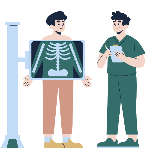
Chest X-ray (CXR)
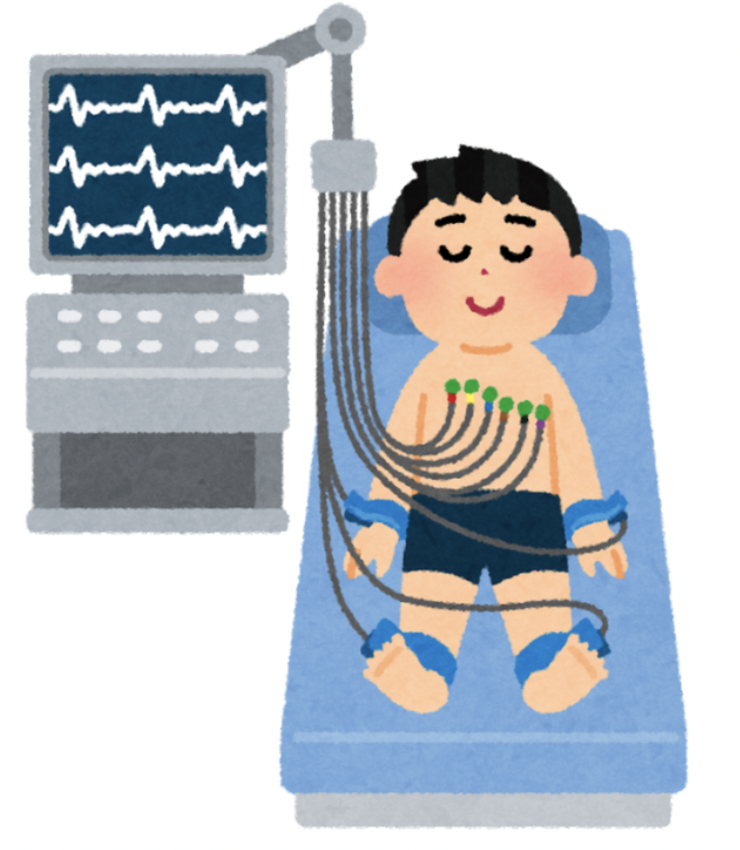
Electrocardiogram (EKG)
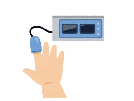
Pulse Oximetry
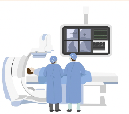
Cardiac Catheterization
Treatments
Soon after birth, prostaglandin is administered through an IV to keep the ductus arteriosus open.5 A stent may also be put in place.3 The stent will create a connection from the aorta to the pulmonary artery by keeping the ductus arteriosus open. By saving this opening, which usually closes after birth, blood can still travel to the lungs and become oxygenated. Another course of treatment is a balloon atrial septostomy. In this procedure, a balloon is placed in the foramen ovale to widen the hole between the two upper heart chambers. This hole increases the amount of blood available to the lungs, but it does allow deoxygenated and oxygenated blood to mix.
Another early treatment option is a balloon valvotomy, a procedure designed to widen a narrowed pulmonary valve3. This is performed by inserting a catheter into a blood vessel and guiding it to the heart. The catheter has a balloon at its tip, which is inflated to enlarge the valve opening. Once the valve is widened, the balloon is deflated and removed
Later, the surgeon may decide to perform a right ventricular outflow surgery to repair and reconstruct the pulmonary valve.5 The connection from the right ventricle to the lungs may be patched with a graft to improve blood flow, as well. A Blalock-Taussig (BT) shunt can also be used in this procedure to increase blood flow to the lungs.
An operation performed later, when the patient is between 4 and 12 months old, is the Bi-directional Glenn procedure. This replaces the BT shunt with a new connection to the pulmonary artery.5 During the procedure, a large vein (vena cava) that returns blood back to the heart is surgically connected to the pulmonary artery instead. This allows blood to move into the lungs to receive oxygen and can help the right ventricle grow. In severe forms of PA/IVS, the Fontan procedure can also be used. Although heart transplants are rarely necessary for children with PA/IVS, they may be considered when no other treatment options are viable.
Additional Resources
References
David M Axelrod, MDStephen J Roth, MD, MPH. Pulmonary atresia with intact ventricular septum (PA/IVS). UpToDate. June 12, 2023. https://www.uptodate.com/contents/pulmonary-atresia-with-intact-ventricular-septum-pa-ivs
Texas Children’s. Pulmonary Atresia with Intact Ventricular Septum (IVS). Texas Children’s. Accessed January 12, 2025. https://www.texaschildrens.org/content/conditions/pulmonary-atresia-with-intact-ventricular-septum-ivs
Mayo Clinic Staff. Pulmonary Atresia. Mayo Clinic. Accessed January 12, 2025. https://www.mayoclinic.org/diseases-conditions/pulmonary-atresia-intact-ventricular-septum/cdc-20396714
The Royal Children’s Hospital Melbourne. Pulmonary Atresia with intact ventricular septum. The Royal Children’s Hospital Melbourne. Accessed January 12, 2025. https://www.rch.org.au/cardiology/heart_defects/Pulmonary_Atresia_with_intact_ventricular_septum/
Boston Children’s Hospital. Pulmonary Atresia. Boston Children’s Hospital. Accessed January 12, 2025. https://www.childrenshospital.org/conditions/pulmonary-atresia